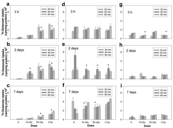
FIG. 3.
Functional assessment of glutamate transporters in isolated neuron (panels a–c) and astrocyte cultures (panels d–f ) and mixed cocultures (panels g–i) after exposure to 1000 MeV/nucleon iron ions. Functional activity was assessed directly by measuring the uptake of 3H-glutamate in cultures 3 h, 2 days and 7 days after irradiation. Data are the average values of three replicate experiments ± SD.

