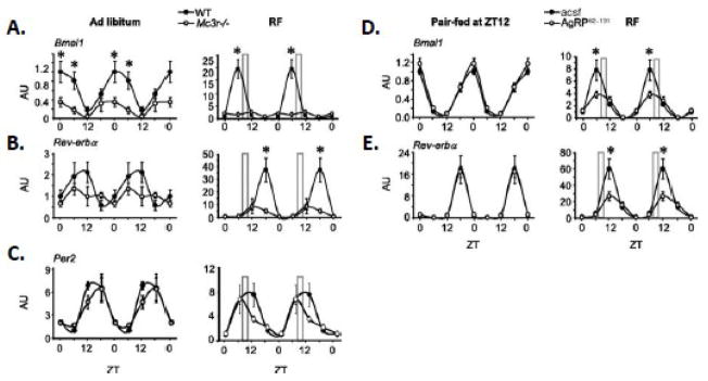
Figure 3.
Mc3r signaling is required for normal liver clock activity during RF. (A–C) Double-plotted expression pattern of Bmal1 (A), Reverbα (B), and Per2 (C) in liver of WT and Mc3r−/− mice fed ad libitum or subjected to restricted feeding (RF, indicated by the grey bar). (D,E) Double-plotted expression pattern of Bmal1 (D) and Rev-erbα (E) in liver of mice treated i.c.v. with AgRP82–131 or acsf pair-fed a nonlimiting amount of food (4.5 g) at ZT12 (6:00 PM, onset of lights off; left panels) or subjected to RF (right panels). *p< 0.05 vs. corresponding WT. AU, arbitrary units. [Adapted from Sutton et al. FASEB J. copyright 2010 with permission from the Federation of American Societies for Experimental Biology]

