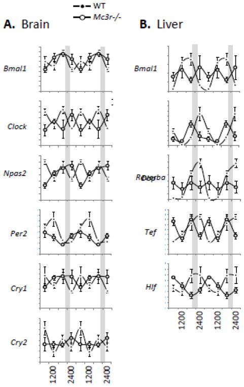
Figure 5.
Arrythmic circadian rhythms of clock gene expression in the brain and liver of Mc3r−/− mice housed in constant dark. Tissues were collected from WT and Mc3r−/− mice at 6h intervals (0600, 1200, 1800 and 2400 h) 4 days after a phase delay in food presentation, and the expression patterns of clock genes assessed in the brain (A) and liver (B) using published methods [81, 83]. The mice were not fed on the day of tissue collection.

