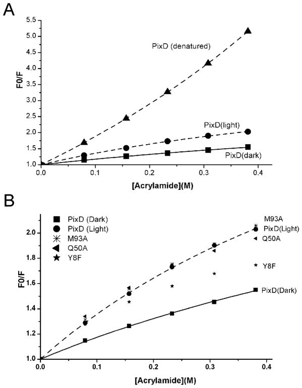
Figure 2.
A: Acrylamide quenching of tryptophan fluorescence of PixD in its native and denatured states. F0 and F are the fluorescence intensity in the absence or presence of various concentrations of acrylamide, respectively. B: Acrylamide quenching of tryptophan fluorescence of PixD and its mutants (Y8F, Q50A and M93A). Only dark sate data are included for the mutants since light state data are essentially the same.

