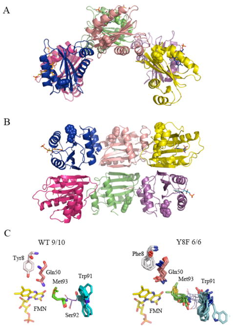
Figure 6.
Crystal structure of the PixD Y8F mutant.. A: Asymmetric unit of Y8F crystal (top view) B: side view. Trp91 side chains are represented as spheres. C: Superposition of Tyr8, Gln 50, Trp91 and Met93 in 9/10 subunits of wild type PixD and of Phe8, Gln50, Trp91 and Met93 in 6 molecules of the Y8F structure. Superposition was performed using Cα of residues 5–140; only amino acids of interest are shown. The FMN molecule was not included in the alignment; however, its position in one molecule is shown for better orientation.

