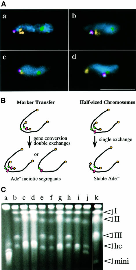
Fig. 6. (A) FISH analysis of wild-type (a) and kms1 mutant (b–d) meiotic prophase nuclei containing the minichromosome ChR33-Tr29. Yellow, rDNA (a, b and d) or telomeres (cos212 in c); green, centromeres (pRS140); red, minichromosome (YIp32). Note that the minichromosome also contains the pRS140 sequence. Nuclei are stained with DAPI (cyan). Scale bar, 10 µm. (B) Experimental designs for the detection of meiotic recombination between a minichromosome and chromosome III (see the text). The rectangles represent the Ade– marker. (C) Pulsed-field gel electrophoresis of stable Ade+ segregants (see the text for details). (Lane a) Chromosomes of S.cerevisiae; (lanes b–j) Ade+ segregants from the mutant cross; (lane k) S.pombe strain containing Ch16 minichromosome. The positions of chromosomes I, II and III, half-sized chromosomes (HC), and minichromosome Ch16 (mini) are indicated on the right hand side.

An official website of the United States government
Here's how you know
Official websites use .gov
A
.gov website belongs to an official
government organization in the United States.
Secure .gov websites use HTTPS
A lock (
) or https:// means you've safely
connected to the .gov website. Share sensitive
information only on official, secure websites.
