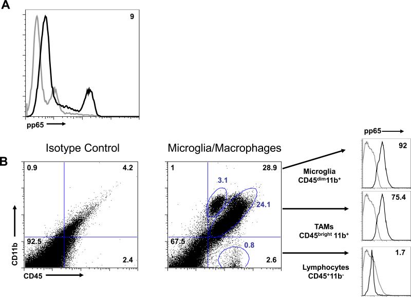
Fig. 1. CMV antigen expression has a tropism for subsets of immune cells within GBMs.
(A) Flow cytometry staining of GBM in single cell suspension shows only a subset of cells are positive for the CMV antigen pp65. The percentage of cells positive is denoted in the upper right quadrant of the histogram. (B) Flow cytometry gating strategy used to distinguish cells of monocyte lineage (such as microglia and MΦs [TAMs]) and lymphocytes based on CD11b and CD45 expression profile and intracellular staining of pp65. Each phenotype was separately analyzed for pp65 expression, as indicated by the histograms. The gray line represents the isotype control, and the black line shows the cells with positive staining for pp65. Percentages of cells are denoted in each quadrant and by their respective gates. The percent positive compared to the isotype control is shown in the right upper quadrant in the pp65 sub analysis panel. This data shown here is a representative of the five newly diagnosed glioblastoma samples analyzed.

