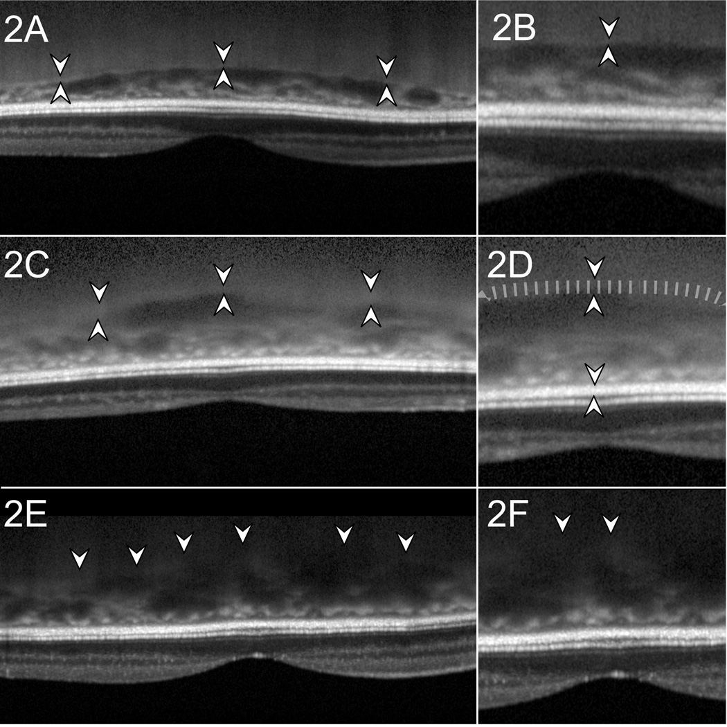
Figure 2.
Examples of scan quality, macular scans
In Figure 2A the choroidal-scleral interface (CSI) indicated by hyper-reflective line between pairs of arrowheads. Figure 2B is a magnified view of 2A and demonstrates what we classified as a good image with a hyper-reflective line whose outer border is marked as the posterior choroidal margin. Figure 2C shows a fair quality image, with thick CSI with lower contrast (between pairs of arrowheads). The CSI was considered thick when it was wider than the thickness of the retinal pigment epithelium (RPE). The outer margin of the CSI was delineated at one RPE thickness behind the inner margin of the CSI. Figure 2d is a magnified image of 2c to illustrate where the outer margin of the CSI was marked (at tip of down-pointing smaller arrowhead). Figure 2e is an example of a poor quality image where the CSI is not distinctly seen. The posterior border of the choroid was marked as a smooth line connecting the most posterior limit of choroidal vascular spaces. Figure 2f is a magnified view of 2e.

