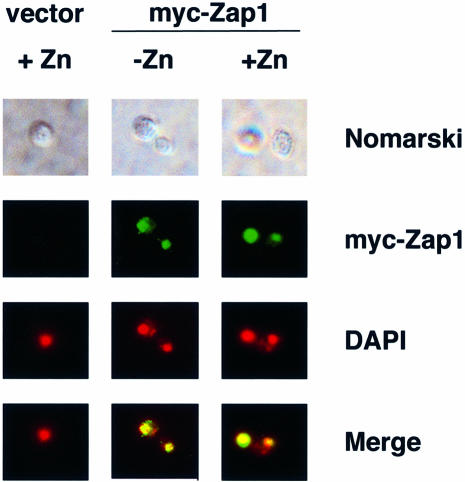
Fig. 2. The subcellular localization of Zap1 is not affected by zinc status. Wild-type (DY1457) cells bearing the vector pYef2 and zap1 mutant (ZHY6) cells bearing pMyc-Zap11–880 were grown to exponential phase in LZM-galactose supplemented with either 5 µM (–Zn) or 1000 µM (+Zn) ZnCl2. Cells were viewed by Nomarski optics or epifluorescence. DAPI was used to stain the nucleus and the myc-Zap1 protein was detected by indirect immunofluorescence. The blue fluorescence of DAPI staining was converted to red and the DAPI and myc-Zap1 images were overlaid using Adobe Photoshop (merge). Yellow color in the merged images indicates colocalization of the markers.

An official website of the United States government
Here's how you know
Official websites use .gov
A
.gov website belongs to an official
government organization in the United States.
Secure .gov websites use HTTPS
A lock (
) or https:// means you've safely
connected to the .gov website. Share sensitive
information only on official, secure websites.
