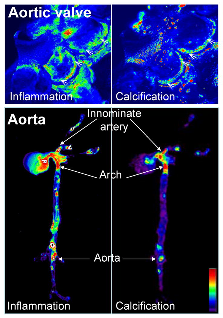
Figure 1. Molecular imaging correlated inflammatory activity, defined as macrophage accumulation, with osteogenesis in the aortic valves and aortas of apoE-/- mice.

Mice were injected with magneto-fluorescent nanoparticles to visualize macrophage accumulation (left panels), and a spectrally distinct bisphosphonate-imaging agent that binds to nanomolar concentrations of hydroxyapatite to detect osteogenic activity (right panels). In the aortic valve (top) and in the aorta (bottom) molecular imaging detected that inflammatory and osteogenic activities colocalized in the areas of highest mechanical stresses at the aortic valve attachment (arrowheads) and at the atherosclerosis prone areas such as innominate artery, aortic arch, and abdominal aorta (arrows). Images were processed using Osirix software. High signal intensities are shown in red-yellow-green. (Adapted with permission from Aikawa E. et al., Circulation 2009 and Aikawa E. et al., Circulation 2007).
