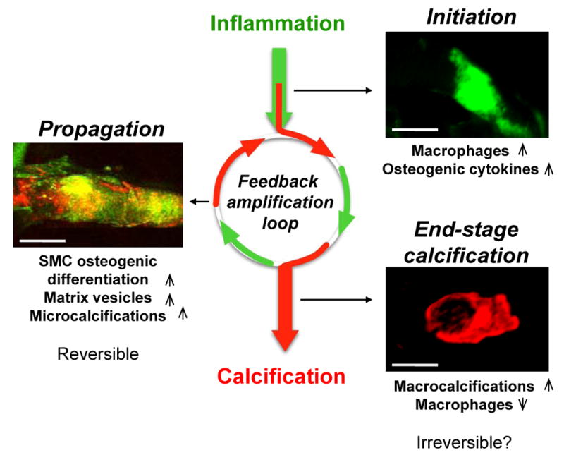
Figure 2. The inflammation-dependent mechanism of calcification visualized by molecular imaging.

Sequential intravital fluorescence microscopy was used to observe changes in inflammation and osteogenesis in atherosclerotic arteries. Two spectrally distinct molecular imaging agents were administered through intravenous injection into apoE−/− mice and the common carotid artery was visualized: magneto-fluorescent nanoparticle to target macrophages (green), and a bisphosphonate-imaging agent to detect osteogenic activity/microcalcifications (red). Three different stages could be identified depending on atheroma progression in 20, 30 and 72 week-old apoE-/- mice feed high-fed diet. In the initiation phase – associated with increased macrophages and pro-osteogenic cytokines – only inflammation was observed (green). In the propagation phase – associated with osteogenic activity and generation of microcalcification – inflammation (green) and calcification (red) overlapped (yellow), suggesting that these two processes develop in parallel. Continued inflammation, in parallel with advancing plaque induces further formation of microcalcifications that provoke additional pro-inflammatory responses from macrophages suggesting that feedback amplification loop of calcification and inflammation drives disease progression. Reducing inflammation through anti-inflammatory therapy at this stage may retard osteogenesis and subsequent calcification. In the end-stage phase – associated with increased mineralization and decreased macrophages – macrocalcifications (red) were observed with limited inflammation. Reversing advanced calcification at this late stage is deemed difficult.
