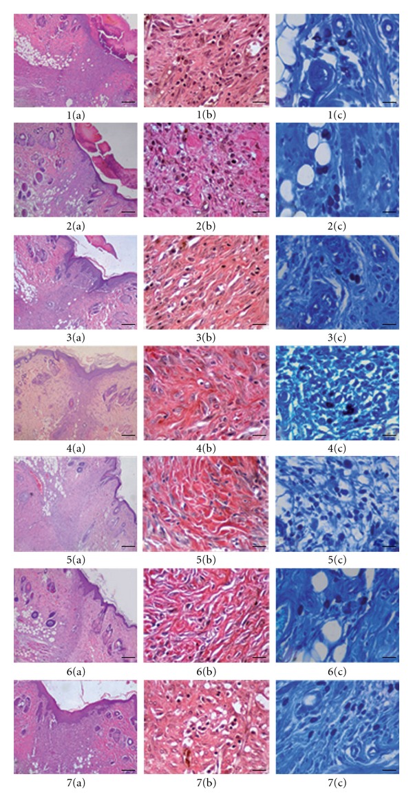
Figure 3.

Histopathological view of wound healing and epidermal/dermal re-modeling in the vehicle, negative control, R. sanctus extracts and Madecassol administered animals. Skin sections show the hematoxylin & eosin (HE) stained epidermis and dermis in (a) and the dermis stained with Van Gieson's (VG) and toluidine blue (TB) in (b) and (c) respectively. The original magnification was 40× and the scale bars represent 25 μm for figures in (a), and the original magnification was 400× and the scale bars represent 100 μm for both (b) and (c). Data are representative of six animal per group. (i) Vehicle group, 10 days old wound tissue treated with only vehicle, (ii) negative c ontrol group (untreated), 10 days old wound tissue (iii) n-hexane extract group, 10 days old wound tissue treated with n-hexane extract, (iv) chloroform extract group, 10 days old wound tissue treated with chloroform extract, (v) ethyl acetate extract group, 10 days old wound tissue treated with ethyl acetate extract, (vi) methanol extract group, 10 days old wound tissue treated with methanol extract and (vi) Madecassol group, 10 day old wound tissue treated with Madecassol.
