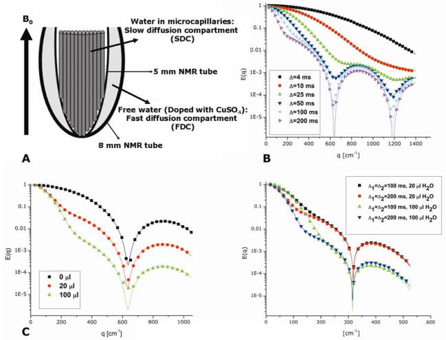
Figure 8.
Single- and double-PFG experiments in the bi-compartmental phantom (81). (A) Cartoon of the bi-compartmental phantom. Microcapillaries are inserted to a 5 mm NMR tube, which is then inserted to an 8 mm NMR tube filled with known quantities of H2O in D2O. Note that the water diffusing freely in the free diffusion compartment (FDC) is completely separated from the microcapillaries (no exchange). The microcapillaries serve as the slow diffusion compartment (SDC) in which restricted diffusion occurs. (B) The s-PFG E(q) plots for varying values of Δ. Note that in the high q-regime, the diffusion-diffraction minima can be gradually observed. At the low q-values, the signal attenuation resembles free diffusion. The experiments were performed on microcapillaries with ID=19±1 μm and with δ=2 ms. (C) E(q) plots for increasing amounts of water in the FDC. The experiments were performed on microcapillaries with ID=19±1 μm and with Δ/δ=100/2 ms. (D) E(q) plots for d-PFG experiments at two different values of diffusion periods and for two different amounts of water in the FDC. The same signal trends were observed as in s-PFG experiments. In all of the plots shown in this figure, the symbols represent experimental data while the solid lines represent the theoretical fittings to the data.

