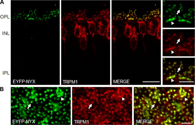
Figure 2.
The TRPM1 channel colocalizes with nyctalopin in DBC dendrites. A, Immunohistochemistry of EYFP-nyctalopin (green) and TRPM1 (red) in retinal cross sections from TgEYFP-NYX mice. The merged images show that expression of TRPM1 in the OPL colocalizes with the EYFP-nyctalopin fusion protein. Scale bar, 10 μm. The OPL at higher magnification is shown in the side panels. Scale bar, 5 μm. B, Whole-mount sections through the OPL of TgEYFP-NYX mice, 5 μm z-stack. EYFP-nyctalopin and TRPM1 are localized to both rod DBC dendrites (arrows) and cone DBC dendrites (arrowheads). OPL, Outer plexiform layer; IPL, inner plexiform layer. Scale bar, 5 μm.

