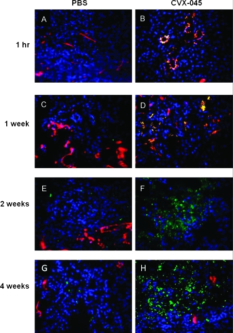
Figure 3.
(A–H) A549 tumors, treated with either PBS or CVX-045 (30 mg/kg 1x/wk), were excised at the time points indicated and sections stained for CD31 (red) to indicate endothelial cells or cleaved caspase 3 (green) to indicate apoptotic cells. Tumor cells were stained with DAPI (blue). Separate images were merged to give yellow regions where red and green overlay, indicating apoptotic endothelial cells.

