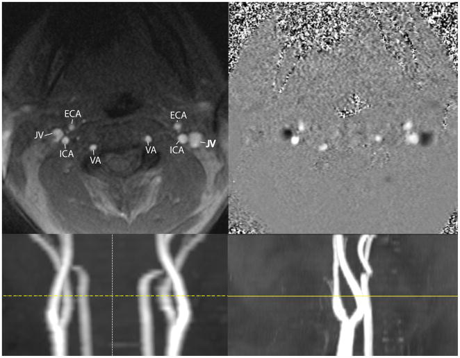
Figure 2.
Upper: Left panel: Magnitude PC-MRA axial single slice (5 mm thickness) demonstrating a typical phase MRA contrast examination. Major vessels are labeled: ECA=external carotid artery, JV=jugular vein, ICA=internal carotid artery, VA=vertebral artery. Image position is above the carotid bulb at the level of the transverse foraminal vertebral artery. Right panel: phase data displayed in gray scale, velocity encoding was 70 cm/sec. Slice location is the same as in the left panel. The jugular veins demonstrate caudal flow, which is encoded as negative values (dark), while the cranial flow is encoded with positive values. Lower: The slice location (yellow line) is depicted on coronal (left) and sagittal (right) vascular scout images.

