
Figure 10. MRI-guided focused ultrasound (MRIgFUS) increases the permeability of the blood–brain barrier (BBB).
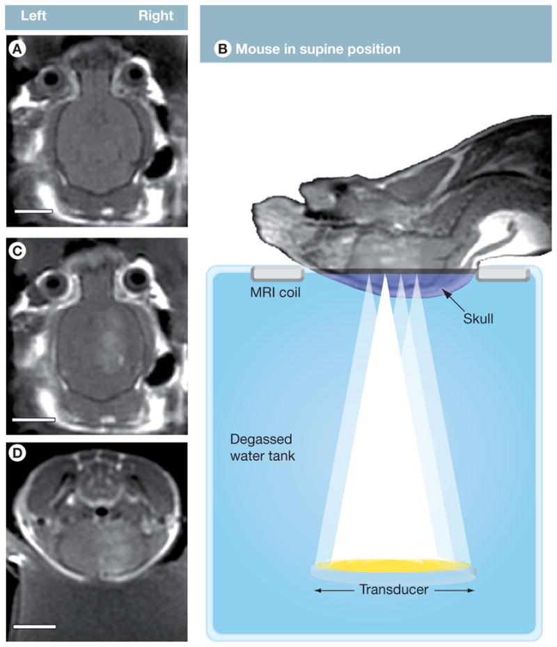
(A) T1-weighted contrast-enhanced MRI scans were used to position ultrasound foci prior to treatment. (B) Mice were positioned in a supine position and injected in the tail vein with microbubbles and gadolinium while US was applied to four aligned spots on the right hemisphere of the brain. Increased BBB permeability was monitored by MRI, visualizing contrast enhancement by the influx of gadolinium (B–D). (A–D: 1286128, TE/TR = 10.4/500.0, FOV = 4 cm, Slice 1 mm, ETL = 4, 3NEX). Scale bars: A, C, D = 0.5 cm.
Reproduced with permission from [98].