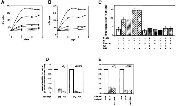
Fig. 1. Effect of androgen and oestradiol on LNCaP cell growth and S-phase entry under different conditions. (A) Quiescent LNCaP cells were left untreated (filled circles) or treated for the indicated times with either 10 nM oestradiol in the absence (open circles) or presence of 10 µM ICI 182,780 (upward filled triangles) or with 10 nM R1881 in the absence (filled squares) or presence (downward filled triangles) of 10 µM Casodex. Control cells were also treated with 10 µM ICI 182,780 (open squares) or 10 µM Casodex (crosses) alone. (B) Cells were left untreated (filled circles) or treated for the indicated times with either 10 nM oestradiol in the absence (open circles) or presence (downward filled triangles) of 10 µM Casodex or with 10 nM R1881 in the absence (filled squares) or presence (upward filled triangles) of 10 µM ICI 182,780. (C) Quiescent LNCaP cells were left unstimulated or stimulated with the indicated compounds for 24 h. BrdU was added and the cells were fixed and permeabilized. DNA synthesis was followed as described in Materials and methods. Several coverslips were analysed and BrdU incorporation calculated according to the formula: percentage BrdU-positive cells = (BrdU-positive cells/total cells) × 100. Results from different experiments were pooled. The means ± SEM are shown. (D) Quiescent LNCaP cells were stimulated for 24 h with either 10 nM oestradiol (right) or 10 nM R1881 (left) in the absence or presence of the indicated inhibitors. PD98059 was utilized at 50 µM and PP1 at 10 µM. BrdU was included in the cell medium and DNA synthesis analysed as described in Materials and methods. Data from different coverslips were pooled and residual BrdU incorporation was expressed as a percentage of steroid-stimulated incorporating cells, which was 28 and 25% for oestradiol- and androgen-stimulated cells, respectively. The basal BrdU incorporation (<5%) was evaluated and subtracted in each case. (E) Quiescent LNCaP cells were either not injected or injected with the indicated constructs (either SrcK– or A221 MEK-1). Either 10 nM oestradiol (left) or 10 nM R1881 (right) was added to the cells for 24 h. BrdU incorporation into DNA was analysed as in (D). Steroid-stimulated BrdU incorporation was 26 and 27% for oestradiol- and androgen-stimulated cells, respectively. The basal BrdU incorporation (<4%) was evaluated and subtracted. For each plasmid, data are derived from at least 150 scored cells. The means ± SEM are shown. A construct expressing the pSG5 empty plasmid was also microinjected into nuclei of quiescent LNCaP cells together with plasmid pEGFP, as an injection marker. The cells injected with pSG5 were left unstimulated or stimulated with either oestradiol or R1881. A total of 23 and 24% of the GFP-expressing LNCaP cells entered S-phase upon stimulation with oestradiol and R1881, respectively. BrdU incorporation of unstimulated cells was <3.5%.

An official website of the United States government
Here's how you know
Official websites use .gov
A
.gov website belongs to an official
government organization in the United States.
Secure .gov websites use HTTPS
A lock (
) or https:// means you've safely
connected to the .gov website. Share sensitive
information only on official, secure websites.
