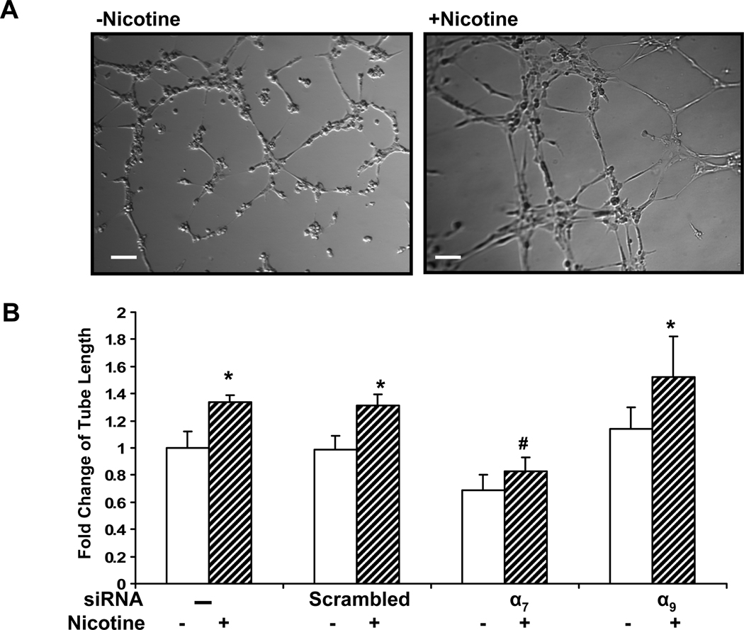
Figure 4. Role of nAChR subtypes in EC tube formation.
(A) Representative phase-contrast micrographs of tube formation of HMVECs on Matrigel. HDMECs were pretreated with siRNA against each individual nAChR subunit for 72 hours. The cells were seeded on matrigel and incubated at 37°C for 6 h in medium with vehicle (left panel) or with nicotine(10−8M, right panel). Bar, 100 µm. (B) Relative fold change in tube length compared to vehicle treated cells. *p<0.05 versus vehicle treated cells. # p<0.05, vs. non-transfected cells exposed to same stimulus.

