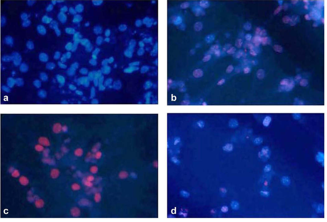
Fig. 3.
Effect of β-AR disruption on doxorubicin-induced cardiomyocyte apoptosis. TUNEL staining of isolated cardiomyocytes from WT and β-AR knockout mice shows a differential susceptibility to doxorubicin. Cells were exposed to doxorubicin 1 µM for 6 h. (a) WT myocytes without doxorubicin (control), (b) WT myocytes with doxorubicin, (c) β2−/− show increased apoptotic nuclei with doxorubicin, (d) β1/β2−/− myocytes show decreased apoptotic nuclei with doxorubicin.

