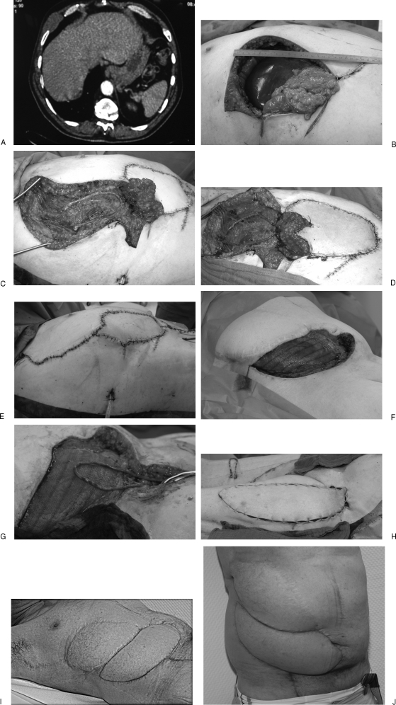
Figure 5.
Case 2: A 54-year-old patient who already underwent sarcoma resection elsewhere. (A) The MRI scan shows recurrence of the tumor (malignant fibrous histiocytoma) at the right lower thorax. (B) After resection of the recurrent tumor, the defect size was 18 × 20 cm. For coverage, a pedicled VRAM flap was planned. (C) The perfusion of the VRAM flap was not sufficient; for salvage of this problem, a free greater saphenous vein graft was anastomosed with the thoracodorsal vessels. (D) The flap is well perfused after anastomosis of the inferior epigastric vessels with the loop. (E) Final result in the operating room. (F) Despite resection with clear margins, another recurrence of the tumor appeared, and the ablative surgeons created a defect of 30 × 15 cm at the lower abdominal wall. Stabilization of the abdominal wall was performed with synthetic mesh. For microsurgical reconstruction, an AV-loop with ipsilateral greater saphenous vein hooked up with the femoral artery was performed. (G) This photograph shows the well-perfused loop. (H) For defect coverage, a combined ALT-TFL flap was harvested. (I) One and one-half years after flap reconstruction, no recurrence of the tumor was seen, and the two free flaps provided a stable thoracic and abdominal wall. (J) The patient is satisfied with the result and is able to perform activities of daily living.

