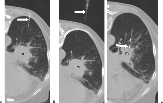
Figure 2.
Needle localization. (A) Computed tomography (CT) image with patient in prone position shows proximal portion of 22-gauge biopsy needle (arrow); left perihilar mass (m). (B) CT image cephalad to A shows needle (arrow) traversing the left posterior chest wall and a portion of the left lower lobe. (C) CT image caudal to A shows needle (arrow) entering the mass (m).

