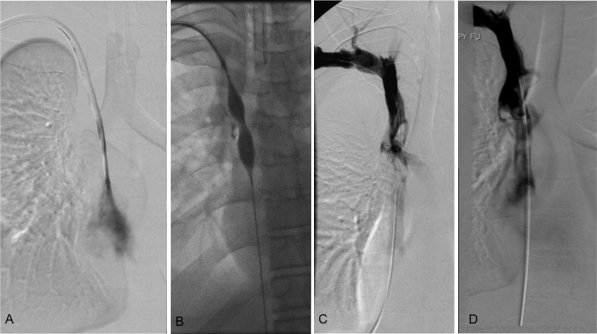
Figure 1.
(A) Digital subtraction venography through a dialysis catheter from a 19-year-old man with bilateral upper extremity as well as facial edema shows the presence of a fibrin sheath. (B) The fibrin sheath was macerated using a 12 mm × 4 cm balloon. (C) Repeat venogram following balloon venoplasty shows a large, acute thrombus in the lower superior vena cava. An infusion catheter was placed across the thrombus and the patient was admitted to the intensive care unit for catheter-directed thrombolysis. (D) Follow-up venogram after a 12-hour infusion of tissue plasminogen activator shows persistent, but decreased clot burden in the superior vena cava. The patient had some immediate postprocedural relief and was started on anticoagulation. Swelling resolved completely upon catheter removal.

