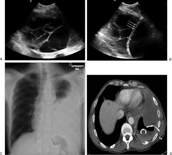
Figure 1.
(A) An ultrasound image shows a multiloculated pleural effusion. (B) A guidewire (triple arrows) is inserted through the initial access needle into the pleural effusion for drainage catheter placement. (C) A chest radiograph shows a large amount of left-sided pleural effusion. (D) An axial computed tomography (CT) image shows large amount of pleural effusion with a 10F pigtail catheter placed percutaneously under CT guidance. The posterior part of the effusion is removed and replaced with air.

