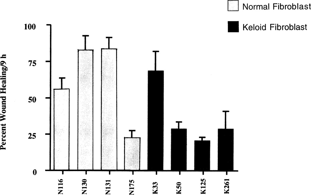
Figure 5.
In vitro wound closure of keloid and normal fibroblasts. Cultures of keloid or normal fibroblasts were cultured in 24 well tissue culture plates. Wounds (400–500 µm) on monolayered keratinocytes were monitored over a period of 24 hours and the area of the wound defect determined using Bioquant software. Fibroblasts from keloids exhibited a wound closure which was slower than that of normal fibroblasts. Values represent mean ± SEM obtained from cultures from cultures of 4 normal and 4 keloid fibroblasts.

