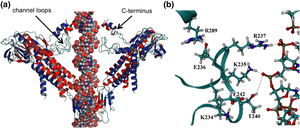
Figure 2.
Modeled structure of the φ29 connector with DNA. (a) The channel loops and C-terminus were modeled into the crystal structure of the connector.11 For clarity, four subunits of the dodecamer are illustrated, including the 1st, 2nd, 8th and 9th monomers. (b) Hydrogen bonds among loop residues (K234-E242), between loop residues (K235, R237, and T240) and DNA, and between the loop and C-terminus (E236-R289).

