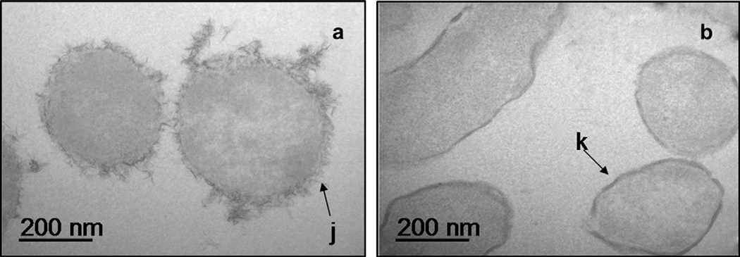
Fig. 11.
Transmission electron micrographs of (a) E. coli with intact LPS (arrow “j”) grown in TSB with 0.5% yeast extract and washed five times with 0.01 M NaClO4 to remove residual media and (b) E. coli after adsorption with 10 mg/L Cu(II) and 5 g/L bacteria. The electron dense region (arrow “k”) reflects Cu accumulation at the outer rim of cells.

