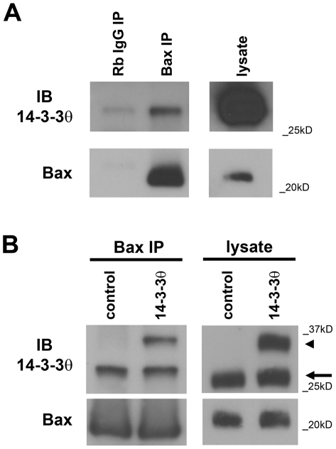
Figure 1. 14-3-3θ immunoprecipitates with Bax in M17 dopaminergic cells.
a) Cell lysates from M17 cells were immunoprecipitated with a polyclonal rabbit antibody against Bax or rabbit IgG, and resulting immunoprecipitants were blotted with a monoclonal mouse antibody against 14-3-3θ in top blot. Lysate lane on right is shown at a different exposure time than the immunoprecipitant lanes from the same gel. Blot was reprobed with anti-Bax antibody to verify Bax pulldown (bottom blot). 14-3-3θ shows specific immunoprecipitation with Bax. b) Cell lysates from M17 cells stably transfected with empty vector or 14-3-3θ tagged with the V5 epitope tag were immunoprecipitated with a polyclonal antibody against Bax and then immunoblotted against 14-3-3θ. Both endogenous 14-3-3θ (lower band marked by arrow) and exogenous, tagged 14-3-3θ (higher band marked by arrowhead) were immunoprecipitated with Bax from cells overexpressing 14-3-3θ, and the total amount of 14-3-3θ immunoprecipitated was increased in 14-3-3θ cells compared to empty vector control cells. Lysate lanes on right were run on a separate gel from the immunoprecipitant lanes. Blot was reprobed with anti-Bax antibody to verify pulldown of Bax (bottom blot).

