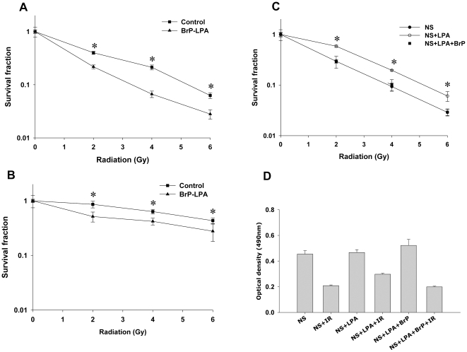
Figure 1. Inhibition of ATX and LPA receptors enhances cell death in irradiated vascular endothelial cells.
(A) HUVEC or (B) bEnd.3 cells were treated with vehicle control (▪) or 5 µM BrP-LPA (▴) for 45 min prior to irradiation. (C) HUVEC were treated with 0.1% fatty acid free BSA (•), 10 µM LPA (○) or 10 µM LPA plus 5 µM BrP-LPA (▪) in serum free medium for 45 min prior to irradiation. The cells were then irradiated with 0, 2, 4 and 6 Gy and plated for clonogenic survival assay. After 2–3 wks, cells were stained with 1% methylene blue and colonies consisting of >50 cells were counted by microscopy. Surviving colonies were normalized for plating efficiency. Shown are average survival fractions and SEM from three experiments; * p<0.05. (D) Equal numbers of HUVEC were plated in 96 well plates and treated with carrier control 3% fatty acid free BSA, 10 µM LPA or 10 µM LPA with 5 µM BrP-LPA in serum free medium for 45 min prior to irradiation. After 96 h, the cell viability was determined using a colorimetric cell proliferation assay (Promega). Shown is the average absorbance at 490 nm with SEM from three experiments; * p<0.05.

