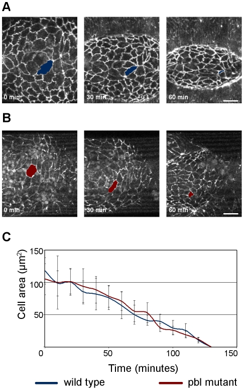
Figure 8. Amnioserosa dynamics in pbl mutants.
(A and B) Frames from movies of arm:GFP-expressing embryos. Dorsal view is shown, scale bars are 20 µm. (A) Wild-type embryo. (B) pbl3/pbl3 mutant embryo. (C) Quantification of amnioserosa cell contraction in a wild-type control embryo (n = 22 cells in 7 embryos) and a pbl3/pbl3 mutant embryo (n = 24 cells in 6 embryos). Bars indicate standard deviation.

