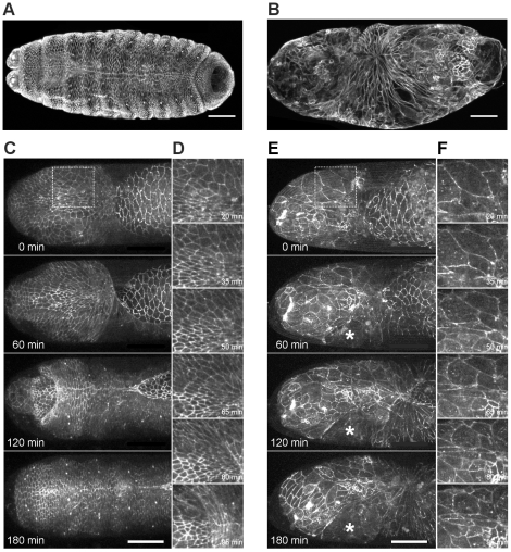
Figure 11. Head involution defects of pbl mutants.
(A and B) Immunofluorescence staining of embryos after head involution stage with anti-Fas3 antibody. (A) Wild-type embryo. (B) pbl3/pbl3 mutant embryo. (C–F) Frames from movies of arm:GFP-expressing embryos. (C) Head region of a homozygous arm:GFP embryo. (D) Enlargement of the boxed region in (C). (E) Head region of a homozygous arm:GFP; pbl3 mutant embryo. Asterisk labels a rip in the head epithelium. (F) Enlargement of the boxed region in (E). (A–F) Dorsal view is shown, scale bars represent 50 µm.

