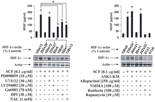
Figure 5. Signalling pathways involved in SCF-induced HIF-1α accumulation in THP-1 human myeloid leukaemia cells.
THP-1 cells were pre-treated for one hour with the indicated concentrations of the outlined inhibitors. In one case cells were transfected with dominant-negative form of ASK1 (ASK1-KM) as indicated in Materials and methods. After that THP-1 cells were exposed for 4 h to 100 ng/ml SCF followed by Western blot analysis of HIF-1α accumulation and ELISA assay of the VEGF release. Quantitative data are mean values ± S.D. of at least three individual experiments. *P<0.01 vs control. a – differences are significant when comparing two indicated values (P<0.01). All Western blot data are from one experiment representative of three that gave similar results.

