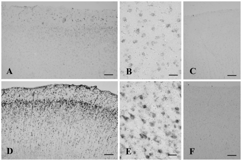
Figure 1. In situ hybridization histochemistry of the cerebral cortex of control (A–C) and Alzheimer's disease (AD) cases (D–F) using antisense (A, B, D, E) and sense (C, F) probes.
Mitochondrial ferritin mRNA localizes mainly in neurons. Both the number and intensity of positive neurons increase in AD cases (D) compared to controls (A). Using sense probes (C and F), no signals are detected in the cortex. Bars = 200 µm in A, C, D, F, and 50 µm in B, E.

