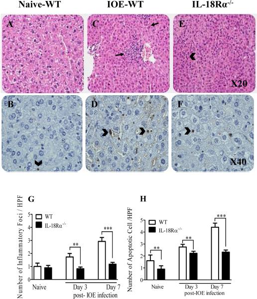
FIGURE 4. Attenuation of liver pathology and cellular infiltration in IOE-infected IL-18Rα-/- mice.
Liver sections from naive (A and B), IOE infected WT mice (C and D), and IOE infected IL-18Rα-/- mice (E and F) harvested on day 7 p.i. are stained with H&E Original magnification x20, or TUNEL x40. H&E staining show that IOE-infected IL-18Rα-/- mice had a lower influx of inflammatory cells (arrows) and a lower number of apoptotic cells (arrowhead) than infected WT and naive mice. TUNEL assays showed substantially decreased number of apoptotic cells (arrowhead) in IL-18Rα-/- mice with ~ 1 to 2 apoptotic cells observed per 40x high power field (HPF) compared with to 5-10 apoptotic cells per 40x HPF in the infected WT mice. Uninfected control mice had only one apoptotic cells/HPF. G and H show the quantitative analysis of the number of inflammatory foci and apoptotic cells determine by H&E and TUNEL assays, respectively, in different groups of mice. The data shown are from a representative mouse from each group (n=4) and are representative of three different experiments. *P <0.05, **P< 0.01. , ***P< 0.001.

