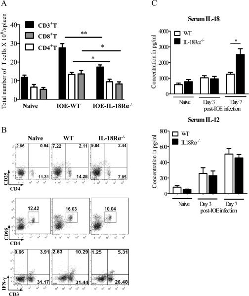
FIGURE 7. IL-18/IL18Rα interaction is essential for the expansion and activation of type-1 cells during Ehrlichia infection.
Splenocytes harvested from naive, IOE-infected IL-18Rα-/- and WT mice on day 7 p.i. were stimulated with IOE Ags. Lymphocyte population was gated based on forward and side scatter and analyzed by flow cytometry. A, IOE infection in IL-18Rα-/- mice resulted in a decrease in the absolute numbers of total CD3+ T cells, CD4+CD3+ cells (CD4+ T cells), and CD8+CD3+ cells (CD8+ T cells) compared to infected WT mice. B, Dot plots showing surface expression of activation marker CD25 and apoptotic marker CD95/FAS, respectively, on CD4+ lymphocytes as well as intracellular IFN-γ production by CD3+ T cells (type-1 T cells). C, Serum cytokine levels of IL-18 and IL-12 in naive, IOE- infected WT and IL-18Rα-/- mice on day 3 and 7 p.i. The data shown are from a representative mouse from each group (n=4), and the numbers indicate the percentage of cells within each quadrant. The data shown are representative of three different experiments. . *P <0.05, **P< 0.01.

