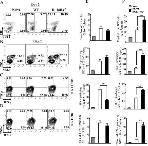
FIGURE 8. Enhanced resistance to Ehrlichia infection in IL-18Rα-/- mice is associated with enhanced expansion of NKT cells and altered function.
WT C57BL/6 and IL-18Rα-/- mice were infected with a high dose of IOE. Splenocytes were harvested from naive, IOE-infected IL-18Rα-/- and WT mice on day 3 (A) and 7 (B-F) p.i., were stimulated in vitro with IOE Ags followed by intracellular cytokine staining. A and B, dot plots showing an increased percentage of CD3+NK1.1+ NKT cells, but not CD3-NK1.1+NK cells, on days 3 and 7 p.i., respectively, in IL-18Rα-/- mice compared to infected WT controls. C and D, cells were gated on NKT and NK cells, respectively, and analyzed for IFN-γ and TNF-α production. E and F show the absolute number of IFN-γ only, TNF-α only and IFN-γ and TNF-α producing NK (E), NKT (F) cells in the spleens of naive, IOE-infected WT and IOE-infected IL-18Rα-/- mice. Data shown are from a representative mouse from each group (n=4), and the numbers indicate the percentage of cells within each quadrant. The data shown are from one experiment that is representative of three different experiments. **P < 0.01. ***P < 0.001.

