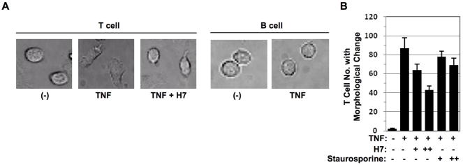
Figure 1. TNF induced T cell morphological change is serine phosphorylation dependent.
(A) Peripheral T cells and peripheral B cells obtained from healthy blood donors were cultured in the chambers coated with ICAM-1 in the presence or absence of TNF (200 ng/ml) with or without serine kinase inhibitor H7 pretreatment for 1hr and immediately subjected to time-lapse fluorescence imaging at 37°C. Cell morphological changes were recorded at time 0 or 30 min after. (B) Peripheral T cells and peripheral B cells obtained from healthy blood donors were cultured in the chambers coated with ICAM-1 in the presence or absence of TNF (50 ng/ml) with or without different dose of serine kinase inhibitors H7 or staurosporine pretreatment for 1 hr and immediately subjected to time-lapse fluorescence imaging at 37°C. Cell numbers with morphological changes were counted after 30 min. Data presented are mean ± SD. of triplicate determinations.

