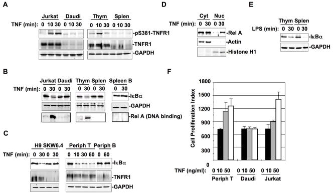
Figure 5. TNF activates NF-κB in T cells via TNFR1-S381 phosphorylation and TNFR1-TRADD complex formation.
(A) Jurkat cells, Daudi cells, mouse thymocytes, and mouse splenocytes were treated with TNF (50 ng/ml) for different times as indicated. Whole cell extracts (equal amount) were subjected to Western blot analysis with anti-pS381-TNFR1 or anti-TNFR1. (B) Jurkat cells, Daudi cells, mouse thymocytes, mouse splenocytes, and purified mouse spleen B cells were treated with or without TNF for 30 min. While the cytosolic fractions prepared from these cells were analyzed with anti-IκBα in Western blot (upper panel), the nuclear fractions were incubated with κB-site oligo-agarose beads overnight at 4°C. DNA precipitated proteins were subjected to Western blot analysis with anti-RelA (low panel). (C) Whole extracts from H9 and SKW6.4 cells treated with or without TNF (50 ng/ml) for 30 min were analyzed with anti-IκBα or anti-TNFR1 in Western blot. Human peripheral T and B cells from normal blood donors were treated with TNF for different times as indicated. Whole cell lysates prepared from these peripheral T or B cells were analyzed with anti-IκBα or anti-TNFR1 in Western blot. (D) Cytosolic and nuclear fractions from above human peripheral T cells treated with or without TNF for 30 min were analyzed for Rel-A nuclear translocation in Western blot. (E) Whole cell extracts of mouse thymocytes or splenocytes treated with or without LPS (10 ng/ml) for 30 mins were analyzed with anti-IκBα in Western blot. (F) Equal amount (4×10⁵) of peripheral T cells, Daudi cells, and Jurkat cells were incubated with or without TNF (10 or 50 ng/ml) for 24 hrs in 96-well plate. Cell proliferation was determined by applying CyQUANT cell proliferation assay kit from Invitrogen (Carlsbad, CA) according to the protocol. Cells were lysed by addition of 100 μl CyQUANT GR dye buffer and the fluorescence intensity was measured with a microplate fluorescence reader FLx800 from Bio-Tek Instruments (Winooski, VT) with excitation at 485nm and emission detection at 530 nm. The fluorescence intensity is presented as cell proliferation index.

