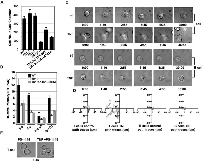
Figure 6. TNF induces T but not B cell migration.
(A) Equal amount (5×10⁵) of mouse embryonic fibroblasts obtained from TNFR1−/−, TNFR2−/− or TNFR1 and TNFR2 double−/− (TNFR1,2−/−) mice, as well as the TNFR1-WT or TNFR1-S381A reconstituted fibroblasts were seeded in the upper chamber (polycarbonate membranes with 8.0-μm pore size) in DMEM medium supplied with 5% FBS. 12 hrs later, the cells migrated to the low chamber were fixed, stained with H&E, and visualized with a light microscope according to our published protocol (13). The above H&E stained cells in the lower chamber were counted from three vision areas with a light microscope. (B) In TNFR1−/− MEFs or TNFR1, TNFR2−/− MEFs with TNFR1-S381A mutant reintroduction, indicated genes were analyzed for their transcriptional regulation by performing RT-PCR. Data are expressed as mean ± S.D. of three experiments. (C) Peripheral T cells and peripheral B cells obtained from healthy blood donors were cultured in the chambers coated with ICAM-1, in the presence or absence of TNF (200 ng/ml) and immediately subjected to time-lapse fluorescence imaging at 37 °C. Cell morphological changes were recorded at different times (min) as indicated. Experiments were repeated on T lymphocyte preparations from three independent donors. (D) The migration of the above T and B cells in (C) was tracked over a 30-min period. Each line represents one cell. (E) TNF treatment at indicated time failed to induce cell morphological change in peripheral T cells pretreated with IKKβ inhibitor PS-1145 for 2 hrs.

