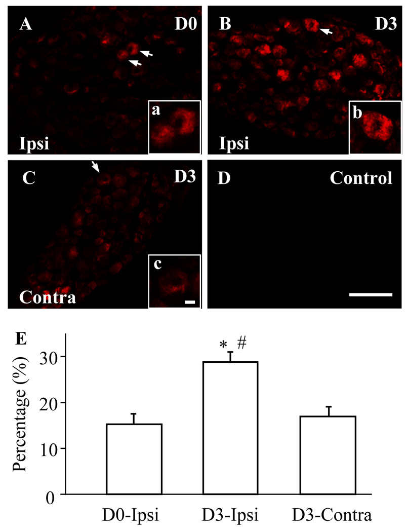
Fig. 2.
Spinal nerve ligation (SNL) increased the number of Nav1.1-positive neurons in the L5 dorsal root ganglia (DRG). A–C: DRG sections were immunostained with anti-Nav1.1. Representative images of naive DRG (A), ipsilateral (Ipsi) L5 DRG (B), and contralateral (Contra) L5 DRG (C) on day 3 post-SNL. Insets a–c are high magnification images of the of Nav1.1-positive cells indicated by the arrows in A–C, respectively. D: A negative control showing absence of signal in the DRG when primary antibody was omitted. Scale bars: 200 µm in A–D and 20 µm in a–c. E: Statistical summary showing the percentage of Nav1.1-positive neurons in naïve (D0) DRG, ipsilateral L5 DRG (B), and contralateral L5 DRG (C) on day 3 (D3) post-SNL. * P < 0.05 vs the corresponding naive group. # P < 0.05 vs the corresponding contralateral side.

