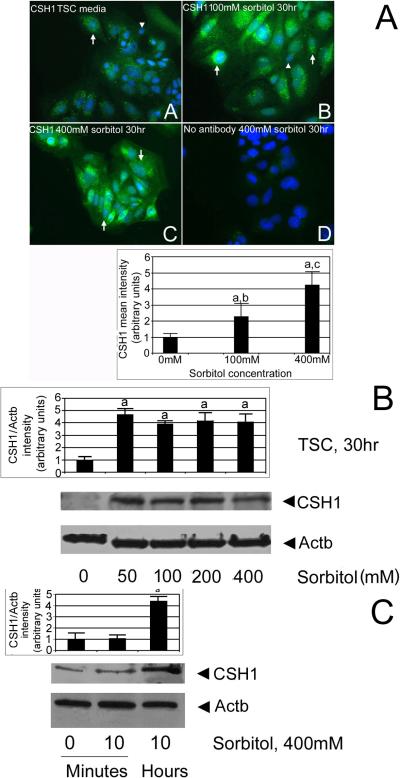
Figure 6.
CSH1 protein is induced in nearly all stressed TSCs in a time- and dose-dependent manner. (A) CSH1 protein is induced in cultured TSCs by 100 or 400mM sorbitol. TSCs were cultured in 0 (A) 100mM (B), or 400mM (C) sorbitol for 30hr and then fixed and stained for CSH1 using indirect immunocytochemistry. In (D), 400mM sorbitol-treated TSCs were probed without primary antibody. Triplicate biological experiments indicate that (a) stress at 100 or 400mM sorbitol induced significant amounts of CSH1 (post hoc Dunnett t test shows p<0.01), (b, c) 100 and 400 were significantly different than each other (post hoc Dunnett t test shows p<0.01). (B). CSH1 protein is induced in cultured TSCs by 50–400mM sorbitol after 30hr. TSCs were cultured in 0, 50, 100, 200, or 400mM sorbitol for 30hr and lysates were fractionated by SDS-PAGE, blotted, and stained for CSH1 and Actb. In (D), 400mM sorbitol treated TSCs were probed without primary antibody. Triplicate biological experiments indicate that stress induced significant amounts of CSH1 (a) at 50–400mM sorbitol (post hoc Dunnett t test shows p<0.01). (C) CSH1 protein is induced in cultured TSC by 10hr, but not by 10minutes. TSCs were cultured in 400mM sorbitol for 10minute or 10hr, and lysates were fractionated by SDS-PAGE, blotted, and stained for CSH1 and Actb. Triplicate biological experiments indicate that stress induced significant amounts of CSH1 (a) at 10hr post hoc Dunnett t test shows p<0.01). In all panels, flags show standard error mean across replicates.

