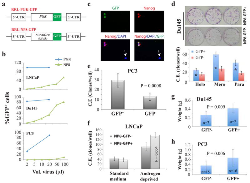
Figure 2. NANOGP8-expressing PCa cells possess CSC properties.

a) Schematic of RRL-GFP lentiviral reporter constructs. GFP, green fluorescence protein; PGK, phosphoglycerate kinase; NP8, NANOGP8 promoter. b) FACS analysis of the percentage of GFP+ cells following transduction with the promoter reporter lentiviruses. 50K cells/well in 12-well dishes plated 1-day earlier were transduced with the indicated volumes of lentivirus. Cultured cells were maintained in fresh media and passaged until FACS analysis ∼ 5 d post-transduction. c) Immunostaining of NP8-GFP transduced LNCaP cells for NANOG (red) demonstrates co-localization of GFP and NANOG expression. Original magnifications, x200. (d-f) FACS purified viable (7AAD-) NP8-GFP+ PCa cells transduced (7-10 d prior) with the NP8-GFP lentivirus exhibit enhanced cloning efficiency (C.E.). d) NP8-Du145 cells (200 cells/well in 6-well culture plate) scored 14 d post-plating: holo, holoclones; mero, meroclones; para, paraclones. *P <0.001. e) NP8-PC3 cells (200 cells/well in 6-well culture plate) scored 14 d post-plating. f) LNCaP cells assayed in parallel at clonal density in androgen-deprived conditions (CDSS + 20 μM bicalutamide) versus standard growth media (2K cells/well and 200 cells/well, respectively) scored 14 d post-plating. NP8-GFP+ LNCaP cells displayed increased C.E. only in androgen-deprivation conditions. g-h) NP8-GFP+ PCa cells are more tumorigenic than NP8-GFP- cells. FACS-purified GFP+/- cells were injected s.c in Matrigel in NOD/SCID-γ recipients. In g), 5K each of NP8-GFP+/- Du145 cells were injected and tumors harvested at ∼2 months. In h), 1K each of purified NP8+/- PC3 cells were injected s.c in the 1° and 2° tumor transplantation experiments (harvested at d 56 and day 49, respectively). Shown are the pooled data.
