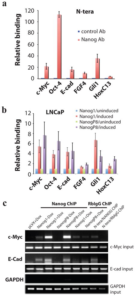
Figure 4. Overexpressed NANOG1 and NANOGP8 proteins in cancer cells bind the expected molecular targets.

a) ChIP assays in N-TERA human embryonal carcinoma cells. Chromatin prepared from N-TERA cells was used in immunoprecipitations with a Rb polyclonal anti-NANOG Ab or, as control, Rb IgG. DNA co-precipitated in ChIP was then used in PCR analysis of the promoter region of the indicated molecules. The results were expressed as NANOG binding to the targets relative to RbIgG (plotted as 1). b) pLVX-NANOG1/NANOGP8 LNCaP cells (clone D2) were either untreated or induced with dox (500 ng/ml) for 72 h. Chromatin was prepared and ChIP assays were performed similarly to those in N-TERA cells. c) Examples of ChIP assays with GAPDH ChIP as negative control.
