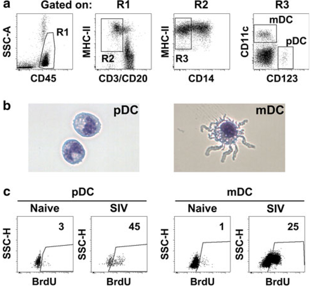
Fig. 1.
Dendritic cell identification in rhesus macaques. a Representative flow cytometry dot plots revealing the gating strategy used to define pDC and mDC within peripheral blood mononuclear cells of an SIV-naïve macaque. b Representative images of pDC and mDC sorted from lymph node cell suspensions of a rhesus macaque. Cells have been stained with modified Wright–Giemsa. c Representative dot plots demonstrating increased blood pDC and mDC incorporation of 5-bromo-2′-deoxyuridine in vivo in acute SIV infection of rhesus macaques relative to pre-infection. Numbers represent percentage of cells in the indicated gate

