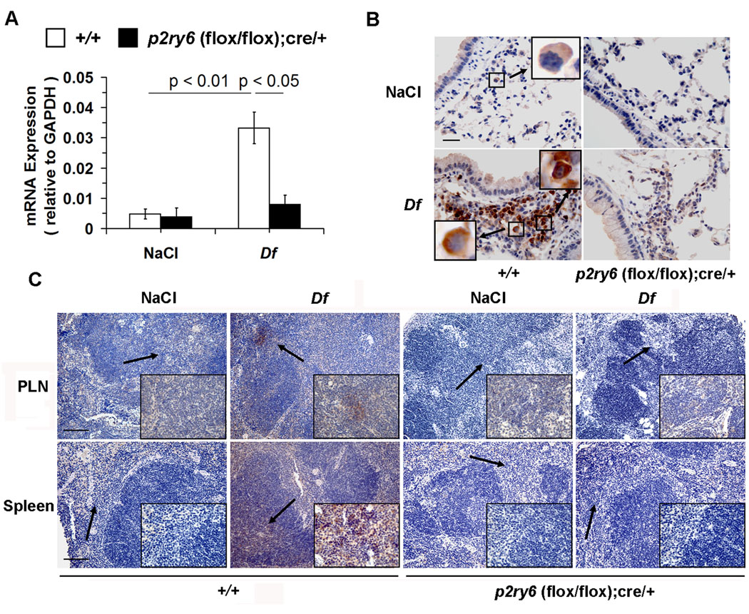
Figure 3. Expression of P2Y6 receptors in the lungs, PLNs and spleen.
(A) qPCR analysis of P2Y6 receptor mRNA in the lungs of NaCl- and Df-treated +/+ (open bars; n = 6 and 18, respectively) and p2ry6 (flox/flox); cre/+ (filled bars; n = 6 and 15) mice.
(B) Immunohistochemical analysis of P2Y6 receptors in the lungs from +/+ and p2ry6 (flox/flox); cre/+ mice exposed to NaCl and Df intranasally. P2Y6 receptor protein, indicated by the brown staining, was detected on cells with the morphology consistent with macrophages (upper left panel, insert) in the lung of +/+ naïve mice and on both macrophages (lower left panel, left insert) and lymphocytes (right insert) in the bronchus-associated lymphoid tissues of +/+ mice. No staining was detected in the lung of NaCl- and Df-treated p2ry6 (flox/flox); cre/+ mice (right panels). Scale bar, 25 µm. (C) Immunohistochemistry of P2Y6 receptors in PLNs and spleen of NaCl- and Df-treated +/+ and p2ry6 (flox/flox); cre/+ mice. P2Y6 receptors were detected on cells located in paracortical T cell-dependent areas of the organs (arrows), as shown in the inserts at higher magnification. Scale bars, 100 µm. Values in (A) are mean ± SEM from three independent experiments. Pictures in (B) and (C) are from one representative mouse per group from one of two independent experiments with similar results. Original magnification, x63 (B), x20 and x63 (C).

