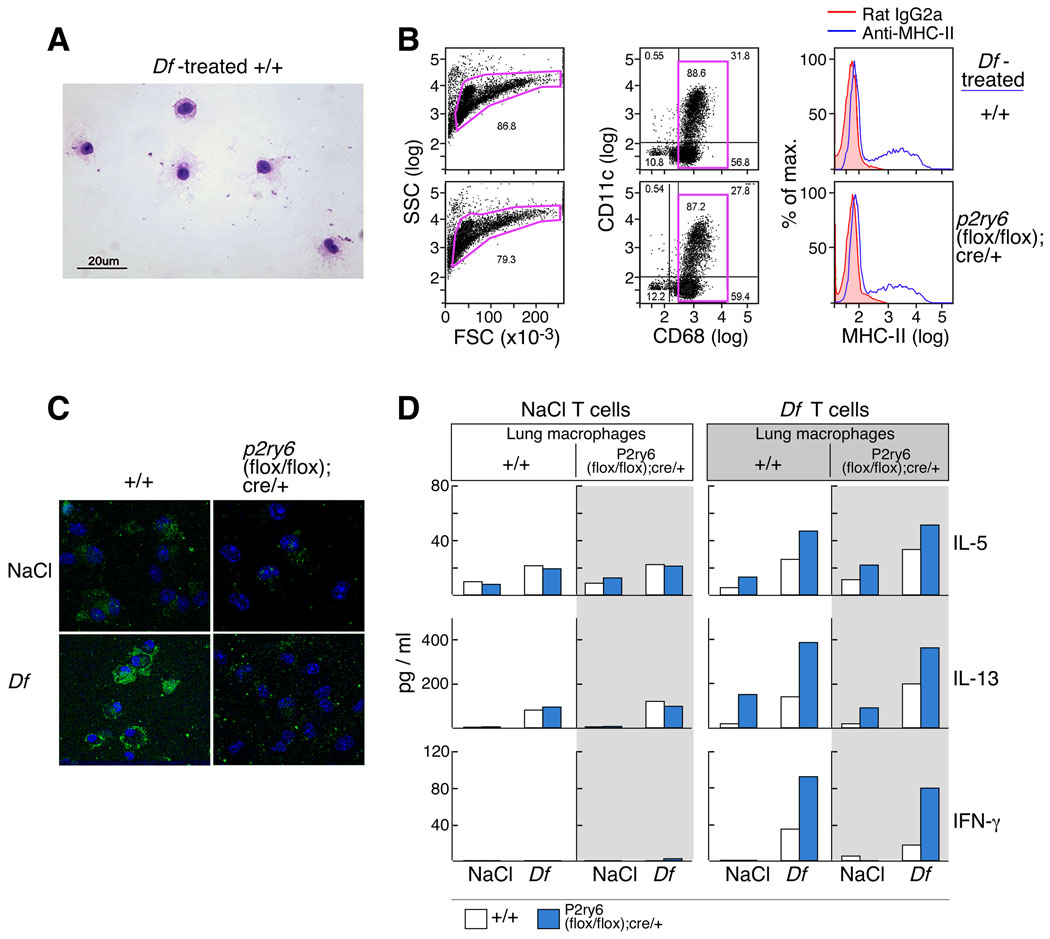
Figure 6. Cytokine release from restimulated cocultures of CD4+ T cells and lung macrophages.
(A) Wrights and Giemsa stain showing enriched lung macrophages from Df-treated +/+ mice. (B) Forward and side scatter characteristics (left), CD68/CD11c staining (middle) and MHC-II staining (right) of the enriched macrophages from the indicated strains used in the co-culture assays. (D) Cytokine release from restimulated cocultures of CD4+ T cells and lung macrophages. Lung macrophages from NaCl- and Df-treated +/+ (unshaded) and p2ry6 (flox/flox); cre/+ (shaded) mice were co-cultured with CD4+ T cells from +/+ (open bars) and p2ry6 (flox/flox); cre/+ (filled bars) mice treated with NaCl (left panels) or Df (right panels). The results are from one experiment, which was repeated twice more with similar trends but with different magnitude of responses.

