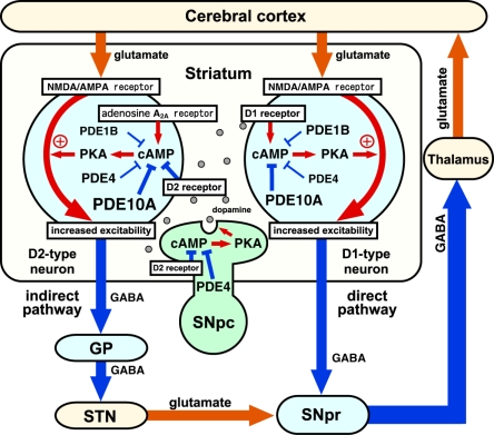
Figure 3.
Roles of phosphodiesterases (PDEs) in the control of basal ganglia–thalamocortical circuitry. Output neurons in the striatum are medium spiny neurons (MSNs), which consist of striatonigral/direct pathway and striatopallidal/indirect pathway neurons. Direct pathway neurons are GABAergic, and inhibit tonically active neurons in globus pallidus interna (GPi)/substantia nigra pars reticulata (SNpr). Indirect pathway neurons are also GABAergic, and activate neurons in GPi/SNpr via inhibition of globus pallidus externa (GPe) GABAergic neurons and activation of subthalamic nucleus (STN) glutamatergic neurons. Direct and indirect pathway neurons induce opposing effects on the output neurons in GPi/SNpr, resulting in dis-inhibition and pro-inhibition of output, respectively, to motor areas of the thalamus and cortex. The inhibition of PDEs increases cAMP/PKA signaling in both direct and indirect pathway neurons. PDE inhibitors that predominantly act in direct pathway neurons work like dopamine D1 receptor agonists and activate motor function, whereas PDE inhibitors that predominantly act in indirect pathway neurons work like dopamine D2 receptor antagonists and inhibit motor function. SNpc, substantia nigra pars compacta. Reproduced with permission from reference Nishi and Snyder (2010).

