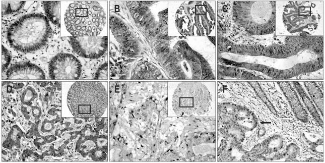
Fig. 1.
Immunostaining of human colorectal cancers with anti-KLF4 antibody (original magnification, ×200). Staining is indicated as a brown precipitate. Unstained nuclei appear blue from the hematoxylin counterstain. (A) Normal appearing colonic mucosa. KLF4 immunostains are predominantly cytoplasmic in the normal crypt epithelium and primarily distributed around the nucleus, a pattern clearly indicative of cell polarity. (B) Colonic villous adenoma. KLF4 immunostains are cytoplasmic and nuclear in the crypt epithelium. Cell polarity is observed. (C) Tubular adenocarcinoma (well-differentiated). KLF4 staining is strong in the cytoplasm and clearly observed in the nuclei of some cells. (D) Papillary adenocarcinoma (moderately differentiated). KLF4 immunostaining is diffusely distributed in the cytoplasm. Several nuclear yellow stains are also observed. (E) Mucous adenocarcinoma (poorly differentiated). KLF4 immunostaining is predominantly cytoplasmic and is weakly positive. Most cells demonstrates a loss of cell polarity. (F) Another specimen illustrates the difference in KLF4 protein between poorly and well-differentiated cells; the yellow stain is darker in the well-differentiated crypt (white arrow) than in the poorly differentiated portion (black arrow).

