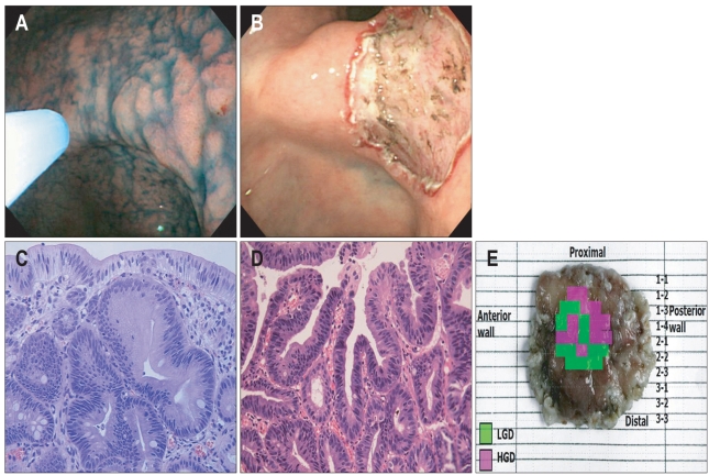
Fig. 1.
A lesion with a histologic upgrade from low-grade dysplasia (LGD) to high-grade dysplasia (HGD) following endoscopic resection. (A) Endoscopic findings of the lesion based on indigo-carmine spray. Endoscopy reveales a 15 mm elevated mucosal lesion with surface nodularity and redness on the posterior wall of the angle. (B) Following endoscopic resection, a 2 cm mucosal defect is observed. (C) Microscopic features of the forceps biopsy. The biopsy specimen shows mild glandular disarray and increased cellularity with basally located, enlarged hyperchromatic nuclei. These findings are consistent with LGD (H&E stain, ×400). (D) Microscopic features of the resected specimen. This portion of the lesion shows marked glandular disarray with vesicular, round nuclei and a marked increase in mitosis. These findings are consistent with HGD (H&E stain, ×400). (E) Map of the resected specimen. The tumor is 15 mm in diameter, and LGD is mixed with HGD.

