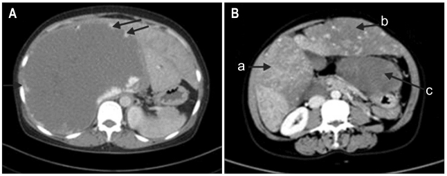
Fig. 2.
(A) Abdominal computed tomography showing a homogenous hypodense lesion with characteristics of a hemangioma (21.9×14.3 cm) in the right lobe. Arrows show the characteristic nodular enhancement. (B) Arrows show three giant hemangiomas. (a) A lesion of 7.8×5.7 cm in S6; (b) A giant hemangioma of 20.2×7.3 cm in left lobe; (c) Another giant hemangioma of 17.7×8.5 cm in the caudate lobe.

